
Is your country {pais}?
From patient registration to sending the images to PACS, on a simulated magnetic resonance scanner. Every finished exam is assessed and recorded for the instructor to follow.
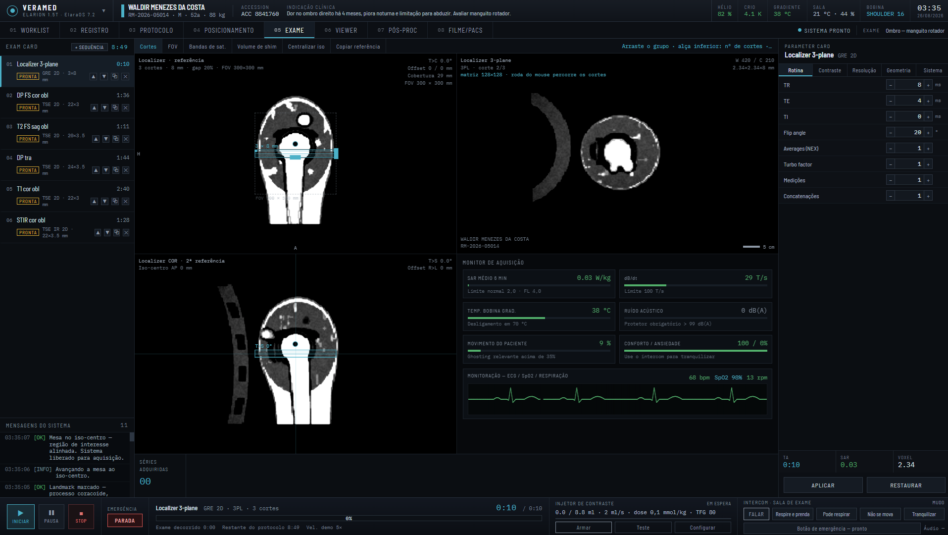
Four materials that complete each other. Each exam report points to what to study again.
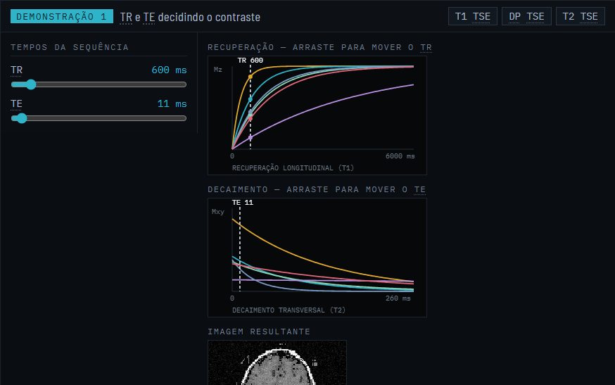
Why each parameter changes the image, with demonstrations where the student moves the value and sees the effect.
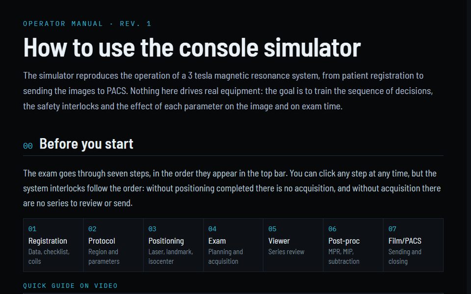
The console workflow step by step, with a video guide for the first session.
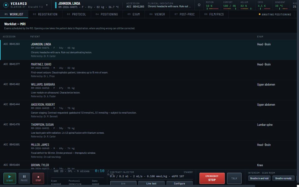
The whole exam, with a patient who moves, tires of breath-holds and squeezes the alarm bulb, and with emergencies.
Twenty-three criteria, each finding with its cause and explanation, and each student's history on the dashboard.
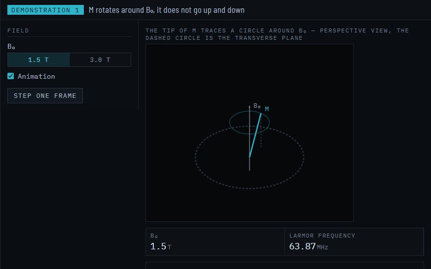
The review and the physics are not running text: each concept has a demonstration with controls, and the result appears right away.
The steps appear in the top bar of the console and can be opened in any order, but the system interlocks follow the sequence: without completed positioning there is no acquisition, and without acquisition there are no series to review or send. The protocol comes before positioning because it determines the coil — on a real console the order arrives through the worklist with the procedure, and the radiographer checks protocol and coil before entering the room.
The criterion is not having the same button. It is that the decision the operator makes here is the one they would make on the scanner, and that the mistake costs the same.
Each of these is explained in detail in the operator manual.
The simulator is faithful in operation, in the planning calculations and in contrast: the sequence of decisions, the safety interlocks, the numbers that appear before you press start and the brightness of each tissue in the image come from formulas with the same proportionalities as the real scanner. What it does not do is reconstruct an exam from the physical signal: k-space is filled from a synthetic image of the slice, not from encoded spins, and the anatomy comes from labelled atlases and procedural models, not from the patient being examined. Below, what stays at each level.
By capability family, against what the four major manufacturers — Siemens, GE, Philips and Canon — offer in common at this level. Product names only appear when the feature is known by them; the comparison is not item by item against any specific scanner.
| Family | Commercial console | The simulator | Verdict |
|---|---|---|---|
| Worklist | DICOM Modality Worklist from the RIS, emergency patient, record merging. | Simulated worklist with accession, indication, referring physician and priority. No emergency patient. | Consistent |
| Protocol tree | Hierarchical tree by region and scanner, site protocols, versioning, dragging sequences between exams. | Flat list of twelve regions with a library of ninety-one programs and positioned insertion in the exam card. | Simplified |
| Graphic prescription | Prescription on three simultaneous localizers, automatic alignment to anatomy (AutoAlign, SmartExam), reference copy between series, tracking. | Prescription on sagittal and coronal localizers, with a check of shift, angulation and coverage against the region reference; geometry copy for 2D sequences. | Consistent |
| Automatic adjustments | Frequency, transmitter gain, receiver gain, higher-order shim, coil element selection. | Automatic frequency, transmitter and volume shim, plus element check; element selection and receiver gain adjustable by hand. No higher-order shim or automatic selection. | Simplified |
| Physiological gating | ECG, peripheral pulse and respiratory belt; trigger delay, acceptance window, arrhythmia rejection, diaphragm navigator. | ECG, peripheral pulse and respiratory navigator, with trigger delay, acceptance window and arrhythmia rejection — a narrow window loses more beats when the rhythm is irregular. No respiratory belt. | Consistent |
| Contrast injector | Volume, flow rate, saline, delay; test bolus and bolus tracking (CARE Bolus, SmartPrep, BolusTrak). | Volume, flow rate, saline, delay, test bolus that measures circulation time, tracking that triggers the series when the contrast arrives, and a block for GFR below 30. | Consistent |
| Real-time acquisition | Adjustment of plane, TR and angle during acquisition; fluoroscopy for manual angiography triggering. | Absent. The acquisition runs to the end or is interrupted. | Gap |
| Reconstruction | Deep learning reconstruction with adjustable denoise level (AIR Recon DL, Deep Resolve, SmartSpeed, AiCE); compressed sensing acceleration. | AI reconstruction level as a parameter, with signal-to-noise gain and apparent sharpness against processing time, and a warning at the high level for smoothing noise aggressively. iPAT as linear acceleration. No real neural network or compressed sensing. | Simplified |
| Post-processing | MPR, MIP, subtraction, ADC maps, perfusion and fusion, with parameters and real results. | The four tasks exist with thickness, increment and rotation parameters, but produce a schematic derived series. | Simplified |
| Viewing | Full DICOM viewer, window presets, hanging protocols, ROI statistics. | Seven tools — window, zoom, pan, distance, ROI, angle and cine — in a configurable grid. | Simplified |
| Sending and documentation | C-STORE to PACS, printing, exam record, structured report. | Simulated transfer to PACS and printer. | Simplified |
| RF safety | Normal, First Level and Second Level mode; B1+rms declared for checking against the MR Conditional implant sheet; nerve stimulation model. | Normal and First Level with a block at 4.0 W/kg; dB/dt with a 100 T/s limit; B1+rms calculated and checked against the implant sheet, with an implant mode that applies the sheet limits. No Second Level. | Consistent |
| Coils | Automatic detection, element selection, coil combination, mixed set-ups. | Nine coils with manual choice and port connection; elements switched on and off with a signal-to-noise penalty for incomplete coverage, and adjustable receiver gain. No automatic detection or combination between different coils. | Simplified |
| Emergencies | Quench button, emergency stop, patient call bell, monitoring. | Five incidents with a step-by-step procedure, drawn during acquisition and with consequences on the system state — plus five additional incidents through the instructor panel. | Goes further |
| Patient behaviour | Does not exist. The patient is real. | Movement, anxiety, comfort, breath-hold tolerance and call bell, evolving with what the operator does. | Goes further |
| Operator assessment | Does not exist. The console does not assess who operates it. | Twenty-three criteria in the closing report, each finding with cause and physical explanation, a log of the session decisions and a performance dashboard with each student's history. | Goes further |
While other MRI simulators pursue image fidelity, ELARION pursues acquisition fidelity: what the operator plans, decides and does during a real exam. The planned geometry changes the slice, the injector and the patient react, and the instructor can trigger incidents.
| What the student trains | ELARION | CorsmedPHYSICS SIMULATION | CLMRIRECORDED SERIES | ScanLabMRPARAMETER LABS |
|---|---|---|---|---|
| The planned geometry changes the image | YesSlices generated from the 3D volume at the planned position, thickness and angle | YesImage simulated from the anatomy, according to the vendor | NoChanging thickness or angle returns the same recorded series | PartialThe page says the student sees the slices the prescription produced |
| Contrast injector | YesLine test, bolus, dose by weight and lockout by GFR | NoNot in the public material | NoNot identified | NoNot on the page |
| Patient and environment that react | YesMotion, anxiety, breath-hold, alarm bulb, ECG and SpO2; quench and cryostat | PartialMotion and artifacts in the image, no live vital signs | No | No |
| The instructor triggers incidents during the exam | YesInstructor panel on a second screen, with incidents triggered live | NoClasses and assessment in an LMS; the instructor does not interfere in the exam | No | NoClass reports and instructor office hours; no intervention in the exam |
| Safety and emergencies | YesSAR, dB/dt, implant sheet and quench with the procedure assessed | PartialSAR calculated at acquisition | NoNo SAR or emergencies identified | PartialSafety quiz before the exam; no SAR or emergencies |
| The whole exam, from RIS to PACS | YesWorklist, registration, positioning, planning, viewer and sending | PartialPatient, planning and protocols; little RIS and PACS | PartialRegistration, queue, viewer and film, no worklist | NoTrains slice planning on its own; registration and PACS do not appear |
| Image formation | PartialContrast from signal equations; aliasing, chemical shift and motion through k-space | YesPhysics simulation from the anatomy, according to the vendor | NoRecorded DICOM series, the same for any parameter | PartialContrast and resolution labs; the page does not say how the image is formed |
Based on the public material of Corsmed (September 2026), ScanLabMR and CLMRI (October 2026). The trademarks belong to their owners.
Every assessed exam goes to the dashboard, and the instructor can trigger incidents during the exam.
Start with the radiology review: it is a prerequisite for the manual and explains the reasons behind the numbers the console asks you to check, in many rich, interactive demonstrations. Then the operator manual, which describes the workflow step by step. Only then the console.
Every closed exam goes into the performance dashboard, where the instructor follows the progress of each student and of the class. The console was designed for a wide screen — from 1480 px wide it appears in full, with no horizontal scrolling — and the room and intercom audio need speakers or headphones.
The console was made for wide screens, from 1480 px, with sound for the intercom. The study material opens on any screen.
Training material. The manufacturer, the equipment, the patients and the exams are fictitious. The simulator does not replace supervised training on real equipment and must not be used for clinical decisions.