
IMAGIX
IMAGIX · MRI SIMULATOR

A magnetic resonance console to train without the scanner

From patient registration to sending the images to PACS, on a simulated magnetic resonance scanner. Every finished exam is assessed and recorded for the instructor to follow.
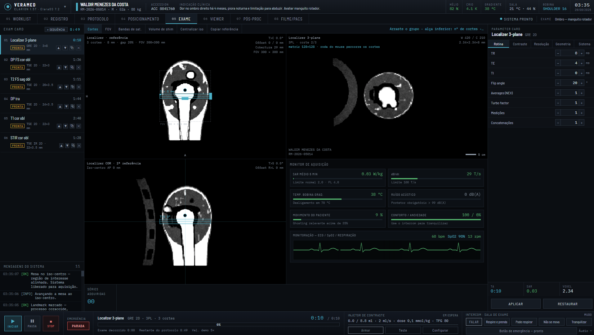
Simulator console at the exam step, on a routine brain: on the left the exam card with the seven sequences of the protocol, in the center the three localizers — sagittal, axial and coronal — with the slice group prescribed over the brain and the lines that mark where each panel is in the other two, in the lower right quadrant the image being acquired, on the right the parameters and the monitor with SAR, dB/dt, acoustic noise and the electrocardiogram trace.
Under a minute: what the simulator does, and how an exam is run on it.
HOW STUDENTS LEARN

Understand, operate, practice and correct

Four materials that complete each other. Each exam report points to what to study again.
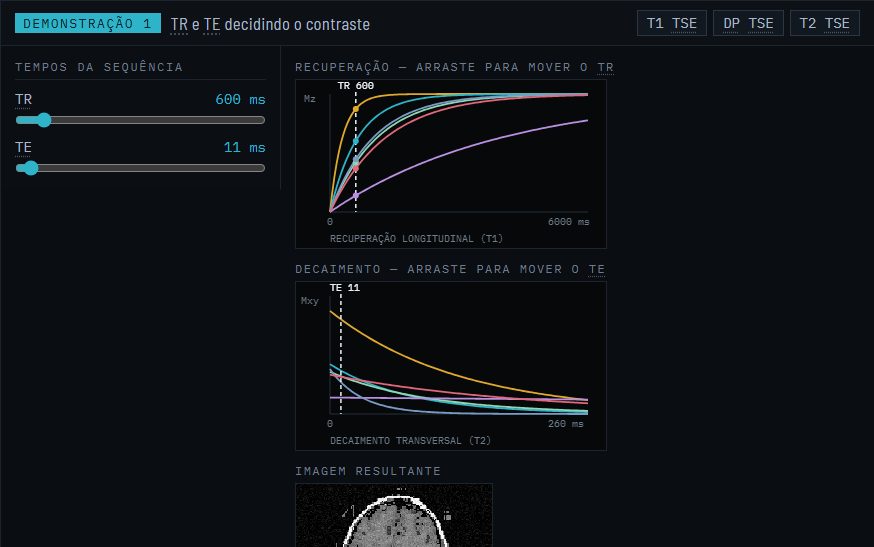
  1. Review demonstration: recovery and decay curves with TR and TE
    1 · UNDERSTAND

    Radiology review and MRI physics

    Why each parameter changes the image, with demonstrations where the student moves the value and sees the effect.
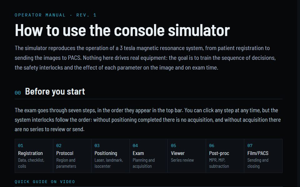
  2. Operator manual with the seven steps of the exam
    2 · OPERATE

    Operator manual

    The console workflow step by step, with a video guide for the first session.
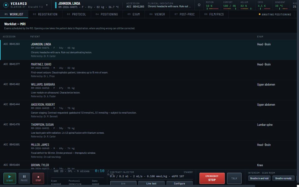
  3. Simulator console on the worklist, with the day's patients
    3 · PRACTICE

    Console

    The whole exam, with a patient who moves, tires of breath-holds and squeezes the alarm bulb, and with emergencies.

  4. 4 · CORRECT

    Exam report

    Twenty-three criteria, each finding with its cause and explanation, and each student's history on the dashboard.

INTERACTIVE DEMONSTRATIONS

Physics the student can move

The review and the physics are not running text: each concept has a demonstration with controls, and the result appears right away.
Review demonstration: recovery and decay curves with TR and TE
TR and TE deciding the contrastDrag TR and TE on the recovery and decay curves and watch the resulting image change from T1 to T2.
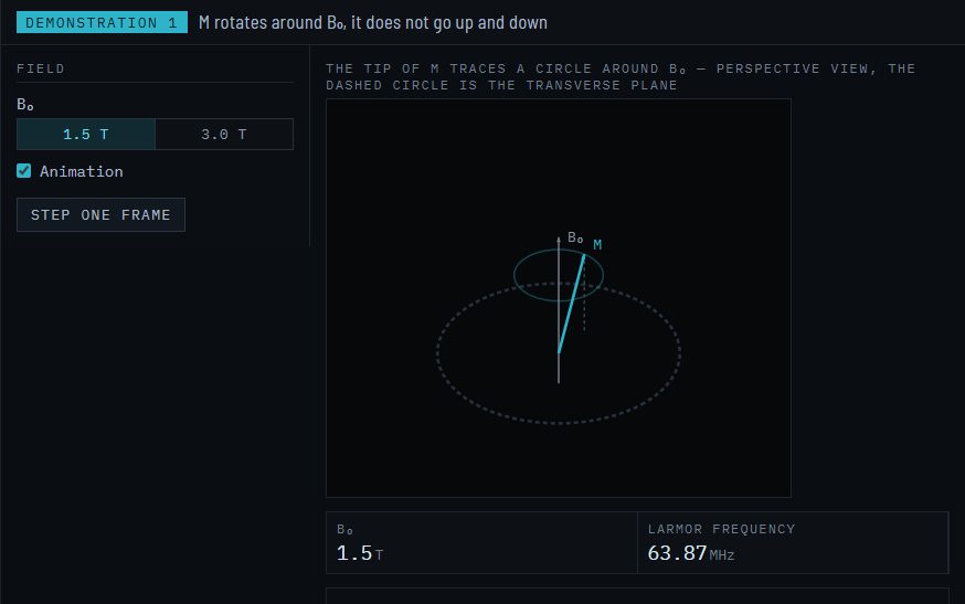
Demonstration: magnetization rotating around the field, with the Larmor frequency
Magnetization rotates around the fieldSwitch 1.5 T to 3.0 T and the Larmor frequency follows.
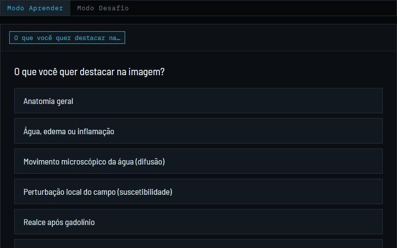
Interactive tree: what you want to highlight in the image
From clinical goal to sequenceThe student starts from what they want to highlight in the image and reaches the sequence family, with the reason for each step.
THE EXAM

The exam, in seven steps

The steps appear in the top bar of the console and can be opened in any order, but the system interlocks follow the sequence: without completed positioning there is no acquisition, and without acquisition there are no series to review or send. The protocol comes before positioning because it determines the coil — on a real console the order arrives through the worklist with the procedure, and the technologist checks protocol and coil before entering the room.

01
Registration
Data, checklist, coils
02
Protocol
Region and parameters
03
Positioning
Laser, landmark, isocenter
04
Exam
Prescription and acquisition
05
Viewer
Series review
06
Post-processing
MPR, MIP, subtraction
07
Film/PACS
Sending and closing
FIDELITY

Compared with a commercial console, aspect by aspect

The criterion is not having the same button. It is that the decision the operator makes here is the one they would make on the scanner, and that the mistake costs the same.

5 consistent 7 simplified 1 gap 3 go further than a real console of 16 aspects compared
The patient responds
A console does not model anxiety or comfort because the patient is on the other side of the glass. Here, not speaking over the intercom has a measurable price in the image, and the emergency call bell is pressed when anxiety crosses a threshold.
The incidents happen
A technologist may go through a whole career without seeing a quench. Here it happens, with the correct procedure required step by step, and the system is left without field afterwards.
The mistake is explained
The final report does not only say what went wrong: it says what would have been right and the physical reason. A console shows the artifact; it does not teach why it appeared.
See the full comparison, line by line

What the simulator keeps track of

Anatomy
Twelve body regions as three-dimensional volumes, with T1, T2 and proton density per tissue.
Patient
Motion, anxiety, comfort and breath change during the exam — and show on the room monitor.
Safety
Checklist before entering, SAR and dB/dt recomputed at every change, and refusal past the limit.
Parameters
TR, TE, matrix, FOV, turbo, acceleration: every change alters time, SAR and the image you get.
Scenarios
Fifteen cases, from a routine head to an acute stroke, each with its own patient and indication.
Emergencies
Five situations with a procedure to follow: ferromagnetic object, alarm bulb, burning, quench and stop.
Instructor panel
A second screen, where the instructor follows the patient signals and triggers incidents.
Report
Twenty-three criteria when the exam is closed, with the mark and what was left undone.

Each of these is explained in detail in the operator manual.

Level of fidelity

The simulator is faithful in operation, in the planning calculations and in contrast: the sequence of decisions, the safety interlocks, the numbers that appear before you press start and the brightness of each tissue in the image come from formulas with the same proportionalities as the real scanner. What it does not do is reconstruct an exam from the physical signal: k-space is filled from a synthetic image of the slice, not from encoded spins, and the anatomy comes from labeled atlases and procedural models, not from the patient being examined. Below, what stays at each level.

CALCULATED
Reacts to each parameter
  • Image contrast, from the signal equations, based on T1, T2, T2*, proton density and diffusion of 25 tissues
  • Geometry: the slice is sampled from a volume, so angling the grid tilts the plane and produces a true oblique slice — the localizer, being an image already acquired, stays still
  • Acquisition time, per sequence and for the whole protocol
  • SAR, with the 2.0 and 4.0 W/kg limits of IEC 60601-2-33
  • Signal-to-noise, voxel size and the effect of field strength
  • TI that nulls fat or CSF, recalculated for the field in use
  • Prescription: centering, angulation and coverage of the slice group
  • Aliasing, susceptibility and motion, each from its own cause
  • Saturation bands and shim volume, with effect and with cost
  • Gadolinium dose by weight and block by GFR
  • Cryostat: helium and magnet temperature rise and fall along a curve, in the quench and in the alarm triggered by the instructor, with recovery
MODELED WITH SIMPLIFICATION
Plausible behavior, not physical
  • dB/dt by a heuristic of bandwidth, thickness and diffusion — without a nerve stimulation model
  • Patient: four variables (movement, anxiety, comfort, breath-hold) with rates per scenario
  • Breath-hold tolerance as a ceiling in seconds, not as respiratory mechanics
  • Motion as amplitude and frequency applied to tissue channels (what breathes, what pulses, the whole body), without diaphragm mechanics or organ deformation
  • Parallel acceleration penalizes signal-to-noise by a heuristic g-factor — it grows with the factor and falls with the number of channels —, without a real parallel reconstruction behind it
  • AI reconstruction gains signal-to-noise and apparent sharpness by a level multiplier (off to high), with a cost in seconds of processing added to the acquisition time — without a neural network or k-space behind it
  • Relaxation times tabulated by field, without variation between individuals
  • Coil elements and channels: manual choice changes signal-to-noise by coverage and combination, without a real spatial sensitivity map behind it
REPRESENTED, NOT SIMULATED
It is on screen, but it is appearance
  • k-space filled from the synthetic image of the slice — matrix, bandwidth, aliasing and motion act on it —, not encoded from spins
  • Anatomy from atlases labeled by region, and from a procedural model where there is no atlas (supine breast, tendon and ligament), not from the patient being examined
  • MPR, MIP and subtraction re-render slices, without reformatting a volume that already exists for that
  • No real DICOM: sending to PACS is a progress bar, not a transfer
  • The room code pairs console and panel, but identifies nobody: who is on the other side is the account the person signed in with
WHERE THE DATA COMES FROM
Protocols
Twelve regions — brain, lumbar spine, knee, shoulder, ankle, wrist, pelvis, prostate, upper abdomen, breast, cardiac and angio — each with its own coil and indication. Changing region reloads the queue and recalculates the total time.
Sequences
Sixty-five sequences with nominal 3 T parameters: 1 mm isotropic sagittal MPRAGE, T2 TSE with an 11-echo train, FLAIR with TI of 2500 ms, DWI EPI b0/1000, SWI, STIR with TI of 180 ms, breath-hold VIBE, TOF. Each one keeps its original values, and it is against them that SAR and time move when you edit.
Library
Ninety-one programs behind the twelve protocols: the sixty-five routine sequences plus twenty-six alternatives, other paths to the same clinical question. STIR and Dixon suppress fat without depending on shim, but cost signal or time; readout-segmented diffusion greatly reduces EPI distortion and takes three to six times longer; a 3D volume does away with the angled group and triples the acquisition; time-of-flight angiography does away with gadolinium and takes almost seven minutes against just over one. The operator adds the program to the exam card in the position they want, as in the protocol tree of a real console, and the sequence already there stays where it was — deleting, skipping or keeping both is the operator's decision, and it is recorded in the session log.
Scenarios
Fifteen, and each one is a set of numbers, not a text: restlessness, breath-hold tolerance, initial anxiety, presence of an implant, GFR and, in two of them, a time ceiling — fifteen minutes in claustrophobia, ten in acute stroke. That is what makes the same protocol behave differently.
Monitoring
Electrocardiogram as a continuous trace, sweeping the screen like a bedside monitor, with P wave, QRS complex and T wave in lead II proportion. Trace, bpm number and beep come from the same beat generator, so in atrial fibrillation the spikes are truly irregular. Next to it, oxygen saturation, which only exists with the patient at isocenter — the oximeter is fiber-optic, because a copper wire inside the magnet forms a loop and burns — and which falls while the breath-hold is sustained.
Cryostat
The quench brings down the field and takes the cryostat with it: helium falls from 82% to zero in twelve seconds and the magnet rises from 4.1 K to over 240 K, with the gauges following live. The gradient coil does not heat up — it works at room temperature with water cooling —, but goes offline with the system; and the room starts to indicate low oxygen, which is the real risk in a quench.
Image
Twenty-five tissues with T1 by field, T2, T2*, proton density and diffusion coefficient, drawn by region and plane. Brightness comes from the signal equations; spectral saturation leaves 12% of residual fat, and noise enters in the proportion of 1 ÷ signal-to-noise.
Artifacts
Seven effects, each with its cause: motion ghosting in the phase direction, aliasing, susceptibility distortion, anatomy outside the slab, fat saturation failure, spike and zipper. Reducing the phase FOV below the body diameter makes aliasing appear in the next image.
Interlocks
Eight-item checklist, coil compatible with the region, table at isocenter, transfer height, pre-scan per sequence and First Level confirmation. Guided mode — on by default — turns the checklist into a requirement to start the acquisition.
Report
The training report is generated from the session log: each finding gives what happened, what would have been right and why, followed by the table of series with the parameters used. The student can export it to hand in to the instructor, and the exam result is recorded in their account — this is what builds the history of the performance dashboard, exam by exam.
WHAT THIS MEANS IN PRACTICE
It is for training operating decisions: choosing the protocol, anticipating the cost of each adjustment, respecting the limits, understanding why a parameter changes contrast and recognizing the artifact by its cause. It is not for validating a clinical protocol, predicting the behavior of a specific scanner or replacing supervised time in the room.

Comparison with commercial consoles

By capability family, against what the four major manufacturers — Siemens, GE, Philips and Canon — offer in common at this level. Product names only appear when the feature is known by them; the comparison is not item by item against any specific scanner.

Family Commercial console The simulator Verdict
Worklist DICOM Modality Worklist from the RIS, emergency patient, record merging. Simulated worklist with accession, indication, referring physician and priority. No emergency patient. Consistent
Protocol tree Hierarchical tree by region and scanner, site protocols, versioning, dragging sequences between exams. Flat list of twelve regions with a library of ninety-one programs and positioned insertion in the exam card. Simplified
Graphic prescription Prescription on three simultaneous localizers, automatic alignment to anatomy (AutoAlign, SmartExam), reference copy between series, tracking. Prescription on sagittal and coronal localizers, with a check of shift, angulation and coverage against the region reference; geometry copy for 2D sequences. Consistent
Automatic adjustments Frequency, transmitter gain, receiver gain, higher-order shim, coil element selection. Automatic frequency, transmitter and volume shim, plus element check; element selection and receiver gain adjustable by hand. No higher-order shim or automatic selection. Simplified
Physiological gating ECG, peripheral pulse and respiratory belt; trigger delay, acceptance window, arrhythmia rejection, diaphragm navigator. ECG, peripheral pulse and respiratory navigator, with trigger delay, acceptance window and arrhythmia rejection — a narrow window loses more beats when the rhythm is irregular. No respiratory belt. Consistent
Contrast injector Volume, flow rate, saline, delay; test bolus and bolus tracking (CARE Bolus, SmartPrep, BolusTrak). Volume, flow rate, saline, delay, test bolus that measures circulation time, tracking that triggers the series when the contrast arrives, and a block for GFR below 30. Consistent
Real-time acquisition Adjustment of plane, TR and angle during acquisition; fluoroscopy for manual angiography triggering. Absent. The acquisition runs to the end or is interrupted. Gap
Reconstruction Deep learning reconstruction with adjustable denoise level (AIR Recon DL, Deep Resolve, SmartSpeed, AiCE); compressed sensing acceleration. AI reconstruction level as a parameter, with signal-to-noise gain and apparent sharpness against processing time, and a warning at the high level for smoothing noise aggressively. iPAT as linear acceleration. No real neural network or compressed sensing. Simplified
Post-processing MPR, MIP, subtraction, ADC maps, perfusion and fusion, with parameters and real results. The four tasks exist with thickness, increment and rotation parameters, but produce a schematic derived series. Simplified
Viewing Full DICOM viewer, window presets, hanging protocols, ROI statistics. Seven tools — window, zoom, pan, distance, ROI, angle and cine — in a configurable grid. Simplified
Sending and documentation C-STORE to PACS, printing, exam record, structured report. Simulated transfer to PACS and printer. Simplified
RF safety Normal, First Level and Second Level mode; B1+rms declared for checking against the MR Conditional implant sheet; nerve stimulation model. Normal and First Level with a block at 4.0 W/kg; dB/dt with a 100 T/s limit; B1+rms calculated and checked against the implant sheet, with an implant mode that applies the sheet limits. No Second Level. Consistent
Coils Automatic detection, element selection, coil combination, mixed set-ups. Nine coils with manual choice and port connection; elements switched on and off with a signal-to-noise penalty for incomplete coverage, and adjustable receiver gain. No automatic detection or combination between different coils. Simplified
Emergencies Quench button, emergency stop, patient call bell, monitoring. Five incidents with a step-by-step procedure, drawn during acquisition and with consequences on the system state — plus five additional incidents through the instructor panel. Goes further
Patient behavior Does not exist. The patient is real. Movement, anxiety, comfort, breath-hold tolerance and call bell, evolving with what the operator does. Goes further
Operator assessment Does not exist. The console does not assess who operates it. Twenty-three criteria in the closing report, each finding with cause and physical explanation, a log of the session decisions and a performance dashboard with each student's history. Goes further
THE CRITERION USED ABOVE
It is not "does it have the button?". It is whether the decision the operator makes here is the same one they would make on the scanner, and whether the price of getting it wrong is the same. A feature can be missing with no harm, if the decision it represents does not exist in the simulated flow; and it can be present and teach nothing, if the mistake costs nothing.
WHAT THIS MEANS
The gaps in the previous section matter to the extent that they reduce the fidelity of the workflow — not because feature parity with a commercial console is missing. The product is in these three rows, not in the ones that are missing.
AMONG SIMULATORS

Compare the solutions

While other MRI simulators pursue image fidelity, ELARION pursues acquisition fidelity: what the operator plans, decides and does during a real exam. The planned geometry changes the slice, the injector and the patient react, and the instructor can trigger incidents.

What the student trains ELARION CorsmedPHYSICS SIMULATION CLMRIRECORDED SERIES ScanLabMRPARAMETER LABS
The planned geometry changes the image YesSlices generated from the 3D volume at the planned position, thickness and angle YesImage simulated from the anatomy, according to the vendor NoChanging thickness or angle returns the same recorded series PartialThe page says the student sees the slices the prescription produced
Contrast injector YesLine test, bolus, dose by weight and lockout by GFR NoNot in the public material NoNot identified NoNot on the page
Patient and environment that react YesMotion, anxiety, breath-hold, alarm bulb, ECG and SpO2; quench and cryostat PartialMotion and artifacts in the image, no live vital signs No No
The instructor triggers incidents during the exam YesInstructor panel on a second screen, with incidents triggered live NoClasses and assessment in an LMS; the instructor does not interfere in the exam No NoClass reports and instructor office hours; no intervention in the exam
Safety and emergencies YesSAR, dB/dt, implant sheet and quench with the procedure assessed PartialSAR calculated at acquisition NoNo SAR or emergencies identified PartialSafety quiz before the exam; no SAR or emergencies
The whole exam, from RIS to PACS YesWorklist, registration, positioning, planning, viewer and sending PartialPatient, planning and protocols; little RIS and PACS PartialRegistration, queue, viewer and film, no worklist NoTrains slice planning on its own; registration and PACS do not appear
Image formation PartialContrast from signal equations; aliasing, chemical shift and motion through k-space YesPhysics simulation from the anatomy, according to the vendor NoRecorded DICOM series, the same for any parameter PartialContrast and resolution labs; the page does not say how the image is formed

Based on the public material of Corsmed (September 2026), ScanLabMR and CLMRI (October 2026). The trademarks belong to their owners.

FOR TEACHERS AND INSTITUTIONS

The instructor follows each student and the class

Every assessed exam goes to the dashboard, and the instructor can trigger incidents during the exam.

How to start

Start with the radiology review: it is a prerequisite for the manual and explains the reasons behind the numbers the console asks you to check, in many rich, interactive demonstrations. Then the operator manual, which describes the workflow step by step. Only then the console.

Every closed exam goes into the performance dashboard, where the instructor follows the progress of each student and of the class. The console was designed for a wide screen — from 1480 px wide it appears in full, with no horizontal scrolling — and the room and intercom audio need speakers or headphones.

FIRST SESSION — SHORT PATH
In the worklist, open the exam of Maria Aparecida (fictional patient), routine brain, tick the whole checklist, confirm the brain protocol, move the table to isocenter, run the pre-scan, acquire the localizer and a T2 sequence, send to PACS and close the exam — it is closing that generates the score and the report. It takes about ten minutes and covers the whole cycle. Then, on the second pass, raise the TE of the T2 and watch the contrast flip, or change the field to 1.5 T in the brand menu and watch SAR drop. The operator manual describes each step in detail.

Runs in the browser, nothing to install

The console was made for wide screens, from 1480 px, with sound for the intercom. The study material opens on any screen.

Notice

Training material. The manufacturer, the equipment, the patients and the exams are fictitious. The simulator does not replace supervised training on real equipment and must not be used for clinical decisions.